Choosing Maxillofacial Implants in 2026 is not a matter of selecting the newest device. It means matching implant design to anatomy, functional goals, and the patient’s medical history. A titanium plate may support a reconstructed jaw, while a custom implant may address a specific facial contour. Neither choice is right for every case.
Dr. Peter D. Waite is an oral and maxillofacial surgeon whose clinical expertise is relevant to this field. To avoid presenting an unverified quotation as fact, the following is a paraphrase, not his verbatim wording: implant decisions should prioritize function, safety, and the needs of the individual patient. That principle matters in the clinic. A scan can show bone volume, but it cannot capture every concern a patient brings to a consultation.
This guide explores the details that deserve careful review: implant material, fit, fixation, surgical planning, and long-term follow-up. It also considers how a multidisciplinary team can assess risks and explain realistic outcomes. Ask about the evidence behind a recommendation. Ask what could change the plan. Small details matter, including the position of nearby nerves and the condition of the surrounding tissue. The right choice is rarely obvious at first glance. And sometimes, the available evidence leaves room for doubt. A careful decision weighs that uncertainty rather than hiding it.
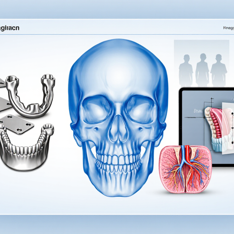
Identify the Defect, Clinical Goal, and Patient Needs
How to Choose Maxillofacial Implants in 2026?
Identify the defect before comparing implant designs. Is the missing structure orbital, nasal, auricular, or part of the jaw? A small orbital defect and a wide maxillary defect create different demands for support, access, and hygiene. CBCT imaging can help clinicians assess available bone and nearby anatomy. No scan tells the whole story. Ask what the patient most wants to regain: facial balance, chewing, speech, or easier prosthesis use.
Patient needs shape the plan as much as anatomy. Consider tissue condition, prior radiation, hand dexterity, cleaning routines, and comfort with follow-up visits. IARC’s GLOBOCAN 2022 estimated 389,846 new lip and oral cavity cancer cases worldwide. That figure is not implant demand; only some patients develop defects requiring reconstruction. Discuss realistic outcomes and limitations with a qualified clinical team. A technically sound plan may still miss the mark if daily care feels too difficult.
Tips: Write down the defect, clinical goal, and patient priority before reviewing options. Ask the patient to describe a typical day. The answer may change the plan.
How to Choose Maxillofacial Implants in 2026? - Identify the Defect, Clinical Goal, and Patient Needs
| Defect or Clinical Situation | Primary Clinical Goal | Common Reconstructive Options | Patient Needs and Selection Factors | Key Considerations |
|---|---|---|---|---|
| Orbital floor or medial-wall defect | Restore orbital support and volume; help address enophthalmos or diplopia when appropriate. | Contourable titanium mesh, porous polyethylene, or composite implants; the choice depends on defect anatomy and surgical plan. | Assess vision, eye movement, globe position, time since injury, associated fractures, and the patient’s functional and cosmetic priorities. | Preoperative imaging and careful positioning are important. Persistent double vision may have causes that an implant alone cannot correct. |
| Cranial or fronto-orbital bone defect | Protect underlying tissues and restore skull contour and, where relevant, orbital symmetry. | Reconstruction may use titanium, patient-specific implants made from materials such as PEEK or PMMA, or bone grafts. | Consider defect size and location, soft-tissue coverage, previous infection or surgery, radiation history, age, and the need for a custom fit. | Material choice is individualized. Infection risk, wound healing, implant exposure, imaging needs, and future procedures should be discussed. |
| Segmental mandibular defect | Restore jaw continuity, facial contour, oral function, and the possibility of future dental rehabilitation. | Depending on the defect and patient, options may include vascularized bone reconstruction, non-vascularized bone grafting in selected cases, and fixation plates as part of a reconstructive plan. | Evaluate defect length and location, remaining teeth, bite, soft tissues, donor-site suitability, overall health, and the patient’s priorities for eating and dental rehabilitation. | A plate can provide fixation or support but does not replace living bone. Larger or complex defects often require multidisciplinary planning. |
| Maxillary or palatal defect | Separate oral and nasal cavities, support speech and swallowing, restore facial form, and plan for dental function. | Options can include a removable obturator, vascularized tissue reconstruction, bone grafting, and dental implants when anatomy and healing permit. | Consider defect extent, remaining dentition, speech and swallowing needs, manual dexterity, ability to maintain an obturator, and treatment preferences. | Prosthetic and surgical approaches may be used separately or together. Dental implant placement depends on available bone, soft tissue, and the overall rehabilitation plan. |
| Facial contour deficiency or asymmetry | Improve skeletal contour or symmetry when the underlying functional structure is stable. | Selected cases may use a patient-specific implant or a surgeon-shaped implant; bone grafting may be considered when appropriate. | Clarify the patient’s appearance goals, asymmetry, skin and soft-tissue thickness, prior operations, and expectations for recovery. | Contour implants are not a substitute for reconstructing a major functional bone defect. Discuss implant visibility, palpability, infection, and revision risks. |
| External ear or nasal prosthesis rehabilitation | Secure a removable facial prosthesis and improve retention and day-to-day usability. | In selected patients, osseointegrated craniofacial implants can retain an external prosthesis; adhesive-retained options may also be considered. | Assess bone availability, skin condition, previous radiation, hygiene capacity, dexterity, prosthesis care, and access to follow-up. | Implant suitability and timing require specialist assessment. The prosthesis and surrounding skin need ongoing cleaning and maintenance. |
| Alveolar bone loss affecting tooth replacement | Restore tooth support and oral function, with or without prior bone augmentation. | Dental implants may be considered after assessment; bone grafting or other augmentation may be needed when bone volume is insufficient. | Consider oral hygiene, gum health, smoking, diabetes control, bone volume, bite forces, and willingness to attend long-term maintenance visits. | Implant placement is not appropriate for every patient or site. A dental and surgical evaluation is needed to assess risks and alternatives. |
Clinical note: This table is for general educational use, not a treatment recommendation. Implant selection should be based on examination, imaging, defect characteristics, and shared planning with the relevant maxillofacial, reconstructive, dental, and prosthetic specialists.
FAQS
Identify the defect’s location and size, such as an orbital, nasal, ear, or jaw defect. Consider available bone, nearby anatomy, and the patient’s main goal.
CBCT imaging can show available bone and nearby structures. It does not tell the whole story, so clinical assessment still matters.
Consider tissue condition, previous radiation, hand dexterity, cleaning routines, and comfort with follow-up visits. Daily care may be harder than expected.
Titanium alloy is strong and often used for load-bearing reconstruction. PEEK is less stiff and may cause less imaging interference. Neither suits every case.
Discuss implant thickness, fixation points, surface texture, and expected loading. Tiny differences can matter near the orbit, nerves, or bite-bearing areas.
A CT-based model can match the defect’s curves and edges. Customization depends on accurate scans and careful manufacturing. Fit may still need adjustment.
Review current safety documents, instructions for use, and lot traceability. Check that sterile packaging is intact and the stated shelf life has not expired.
Confirm authorization for the intended procedure and location. The surgeon should assess anatomy, fixation, instruments, and imaging needs. If documents are vague, pause and ask for clarification.
Conclusion
Choosing Maxillofacial Implants in 2026 begins with understanding the specific defect, the clinical goal, and the patient’s priorities. The right option should support the intended functional or reconstructive outcome while taking comfort, appearance, health history, and individual preferences into account. Careful assessment of the anatomy is essential, and appropriate imaging can help clarify the dimensions, location, and structural requirements of the treatment area.
Next, compare available materials, implant designs, and customization options with the patient’s anatomy and the surgical plan. Confirm that the implant has suitable safety documentation and regulatory status, and that it is compatible with the techniques and equipment required for the procedure. The selection process should also include a practical follow-up plan, with guidance for monitoring healing, assessing the result, and addressing any concerns. Decisions should be made collaboratively by the clinical team and patient, based on current evidence and individual needs.