Choosing the 2026 Best Meniscus Suture Anchor requires more than comparing price, packaging, or headline strength values. Global buyers need evidence that connects laboratory performance with real operating-room demands. A reliable Meniscus Suture Anchor should support secure fixation, controlled deployment, smooth suture handling, and dependable performance across different meniscal tear patterns.
Dr. Robert F. LaPrade has emphasized, “Meniscal preservation should be the goal whenever possible.” That principle still guides responsible product evaluation. Surgeons may work through narrow portals, manage limited visibility, and handle fragile tissue under time pressure. Small design details matter. A sharp needle, predictable anchor insertion, and low-profile fixation can influence the procedure’s efficiency and tissue protection.
However, no anchor is ideal for every patient or technique. Bench-test pullout strength may not predict healing in a vascular red-red zone. A compact device may improve access but provide less familiar handling for some surgeons. These limitations deserve attention, not marketing language. Buyers should review clinical evidence, sterilization records, regulatory documentation, material specifications, and surgeon training support before selecting a supplier.
Global procurement adds another layer. Availability, lot consistency, multilingual instructions, after-sales communication, and regional compliance can affect long-term reliability. The best choice may not be the newest anchor. It may be the one with transparent data, consistent quality, and practical support in the buyer’s market. This guide examines those factors closely, while recognizing that product selection still requires qualified clinical judgment.
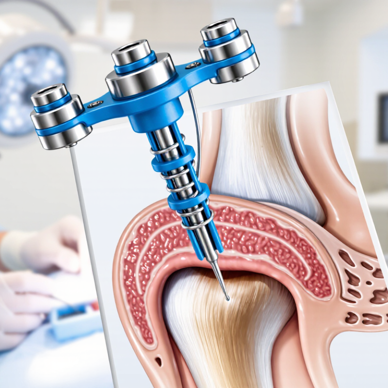
Meniscus Suture Anchors: Purpose, Design, and Clinical Applications
2026 Best Meniscus Suture Anchor for Global Buyers?
Meniscus suture anchors stabilize torn tissue while preserving native knee function. Their design usually combines a small implant, high-strength suture, and a delivery needle. All-inside systems can reduce extra incisions and shorten operative handling. Inside-out repair still offers strong control for accessible tears. Outside-in techniques remain useful near the anterior horn. The best option depends on tear location, tissue quality, surgeon experience, and fixation security.
Clinical evidence requires careful reading. A 2024 systematic review in Arthroscopy reported that meniscus repair failure rates vary widely across techniques and follow-up periods. That variation matters. The implant alone does not determine healing. The American Academy of Orthopaedic Surgeons also emphasizes patient selection, tear pattern, and rehabilitation planning in meniscal care. Market reports from Grand View Research identify continued growth in orthopedic fixation demand, but commercial growth is not proof of superior clinical outcomes. Some product comparisons remain limited. More head-to-head evidence is needed.
Tips: Check anchor deployment depth, suture abrasion, pullout resistance, and MRI compatibility. Match the device to the vascular zone and tear geometry. A compact anchor may simplify access, yet poor placement can damage cartilage. Review regulatory clearance in the target country and confirm training requirements. Document lot information and patient-specific risks. The smallest implant is not always the safest choice.
| Evaluation Dimension | Common Options or Specifications | Clinical Purpose | Key Advantages | Important Limitations | Buyer Selection Considerations |
|---|---|---|---|---|---|
| Primary Function | Fixation of meniscal tissue to the capsule, peripheral rim, or adjacent bone | Stabilizes a repair construct so the meniscus can heal while preserving functional tissue | Supports meniscus preservation instead of routine tissue removal | Healing depends on tear pattern, vascularity, tissue quality, alignment, and rehabilitation | Choose the anchor system according to the intended repair technique and surgeon preference |
| Typical Indications | Peripheral vertical tears, longitudinal tears, bucket-handle tears, selected posterior horn or root-related repairs | Maintains reduction of repairable tears in the vascular or potentially healing meniscal zones | Can be used in arthroscopic meniscal preservation procedures | Not every tear is repairable; chronic, complex, degenerative, or avascular tears may have lower healing potential | Review indications, contraindications, and technique guidance in the applicable instructions for use |
| Anchor Configuration | Small knotless anchor, knotted anchor, all-suture anchor, or suture-loaded anchor | Provides different methods for tissue capture, tensioning, and fixation | Allows the implant to be matched to tear location and access conditions | Each configuration has different handling, pullout, deployment, and revision characteristics | Compare deployment steps, suture management, anchor seating, and compatibility with the repair workflow |
| Anchor Material | Metal, non-absorbable polymer, bioabsorbable polymer, or all-suture construction | Determines the implant’s structural behavior and long-term presence in the body | Material choice can support radiographic visibility, low profile, or gradual resorption | Materials differ in imaging appearance, degradation behavior, inflammatory response, and revision considerations | Confirm material composition, biocompatibility documentation, MRI conditions, and regional regulatory status |
| Suture Type | High-strength braided or tape-style suture; absorbable or non-absorbable options | Captures and compresses the meniscal tissue during healing | High-strength sutures may provide reliable handling and resistance to cyclic loading | Suture cut-through, abrasion, tissue strangulation, or knot-related bulk may occur if technique is inappropriate | Assess suture diameter, tensile performance, abrasion resistance, needle design, and ease of passage |
| Fixation Method | Knotted fixation, knotless tensioning, or anchor deployment after suture passage | Secures the repair at the desired tension and position | Knotless systems can reduce knot prominence and simplify arthroscopic management | Over-tensioning may impair tissue perfusion; inadequate tension may allow gapping or displacement | Prioritize controlled tensioning, tactile feedback, and the ability to verify final fixation |
| Deployment Approach | Arthroscopic insertion through a cannula or portal using a dedicated delivery device | Permits placement in anatomically difficult areas, including the posterior horn | Minimally invasive access may reduce soft-tissue disruption compared with open exposure | Limited visualization, instrument trajectory, neurovascular structures, and cartilage proximity require careful control | Compare inserter length, handle ergonomics, portal compatibility, depth control, and deployment feedback |
| Implant Profile | Low-profile anchor body, compact polymer anchor, or soft all-suture anchor | Reduces prominence within or adjacent to the joint | May be useful where space is limited and where minimizing implant prominence is important | Smaller implants may provide less purchase in poor-quality tissue or weak bone | Balance low profile against required fixation strength and the quality of the meniscal or osseous target |
| All-Suture Anchor Characteristics | Flexible suture-based anchor that expands or bunches within bone after deployment | Provides a small bone fixation point for selected meniscal root or related repairs | Small drill diameter and reduced implant material may be advantageous in limited bone stock | Requires accurate tunnel or pilot-hole preparation and adequate bone quality for deployment | Verify drill size, insertion depth, deployment sequence, pullout data, and compatibility with the surgical technique |
| Bone-Fixation Applications | Selected meniscal root repairs or techniques requiring fixation near the tibial footprint | Restores or stabilizes the meniscal attachment when the repair is directed to bone | Can provide a dedicated fixation point away from the meniscal tissue | Root repair may require additional tunnels, fixation devices, or specialized instrumentation depending on the technique | Assess whether the complete system includes required guides, drills, sutures, and fixation components |
| Soft-Tissue Fixation Applications | Peripheral meniscal repair using capsular or meniscocapsular fixation | Approximates the torn meniscus to the vascular peripheral tissue zone | Can support repair of selected longitudinal or peripheral tears without extensive bone preparation | Fixation quality is highly dependent on tissue capture, anchor placement, and avoidance of excessive tension | Review the recommended tissue thickness, anchor spacing, suture configuration, and access requirements |
| Fixation Strength | Reported as construct or anchor performance under defined laboratory test conditions | Indicates resistance to displacement during early rehabilitation loading | Provides a basis for comparing products when test methods are equivalent | Bench performance does not directly predict clinical outcomes or account for biological healing | Compare test methods, loading direction, cycles, failure mode, bone model, and sample size—not only a single strength value |
| Tissue Compatibility | Materials evaluated for biocompatibility under applicable medical-device standards | Reduces the likelihood of an unacceptable local biological response | Documented material and biological evaluation supports clinical risk assessment | Any implant can be associated with inflammation, infection, allergy, migration, or other adverse events | Request material disclosures, biological evaluation summaries, sterilization information, and adverse-event history where available |
| Imaging and MRI Considerations | MRI-conditional, MRI-compatible under specified conditions, or radiopaque metal components | Determines how postoperative imaging can be performed and interpreted | Appropriate labeling helps clinicians plan follow-up imaging safely | MRI conditions vary by device, field strength, scan parameters, and implant configuration | Check the exact MRI labeling, artifact information, field-strength limits, and local imaging protocols |
| Sterility and Packaging | Single-use sterile device with validated packaging and stated shelf life | Reduces contamination risk and supports operating-room readiness | Preassembled sterile components can simplify preparation | Damaged packaging, expired shelf life, or improper storage can compromise product suitability | Verify sterilization method, packaging integrity, expiration date, storage temperature, and lot traceability |
| Instrument Set Requirements | Dedicated inserter, drill, guide, suture passer, cannula, or tensioning instrument | Enables controlled placement and consistent deployment | A complete instrument workflow may reduce intraoperative delays | Different systems may require proprietary or device-specific instruments | Confirm reusable versus disposable components, sterilization instructions, availability, and replacement-part support |
| Surgeon Handling | Insertion control, suture passage, tensioning feedback, cutting mechanism, and knot management | Influences procedure efficiency and placement accuracy | Predictable handling can shorten learning curves and reduce unnecessary instrument exchanges | Handling preferences vary by surgeon, portal arrangement, tear location, and arthroscopic visibility | Evaluate through structured hands-on training, cadaveric assessment, or supervised clinical use where permitted |
| Potential Complications | Fixation failure, recurrent tear, implant migration, cartilage injury, infection, synovitis, or neurovascular injury | Represents risks requiring prevention, recognition, and management | Risk can be reduced through appropriate indication, accurate placement, and adherence to technique | Complications may occur even when the device is used correctly | Review risk management files, clinical evidence, complaint trends, training materials, and post-market surveillance information |
| Clinical Evidence | Peer-reviewed studies, prospective or retrospective clinical series, biomechanical studies, and post-market data | Supports assessment of safety, healing, reoperation, and functional outcomes | Multiple evidence types provide a broader view of performance | Study results may not be directly comparable because tear patterns, techniques, and rehabilitation protocols differ | Look for evidence relevant to the intended indication, patient population, surgical technique, and target market |
| Regulatory Documentation | Applicable market authorization, technical file, quality-system certification, labeling, and instructions for use | Demonstrates that the device meets the requirements of the target jurisdiction | Clear documentation supports importation, hospital review, and compliant distribution | Approval in one jurisdiction does not automatically authorize sale or use in another | Confirm registration pathway, authorized representative requirements, language labeling, UDI or traceability, and import permits |
| Global Procurement Factors | Lead time, minimum order quantity, cold-chain or storage needs, training support, and service coverage | Determines practical availability for hospitals, distributors, and surgical centers | Reliable supply planning can reduce procedure cancellations and inventory shortages | Customs delays, documentation gaps, and inconsistent component availability may affect continuity | Request a complete product list, lot traceability process, forecast-based supply plan, warranty terms, and technical support contacts |
| Recommended 2026 Selection Priority | Clinical fit, verified safety, handling consistency, regulatory compliance, supply reliability, and total cost of ownership | Creates a balanced basis for comparing generic anchor systems | Reduces the risk of selecting a device based only on price or a single laboratory metric | No single anchor design is optimal for every meniscal tear or every surgical technique | Use a multidisciplinary review involving surgeons, operating-room staff, procurement, quality, regulatory, and biomedical engineering teams |
FAQS
It stabilizes torn meniscal tissue and supports preservation of normal knee function. The implant, suture, and delivery system work together.
The choice depends on tear location, tissue quality, anatomy, and surgeon experience. All-inside, inside-out, and outside-in methods each have practical uses.
No. They may reduce extra incisions and handling time, but placement still requires careful control. A small device is not automatically safer.
Review pullout resistance, deployment depth, suture durability, and relevant test models. Clinical failure rates vary across techniques and follow-up periods. Evidence remains uneven.
Consider tear geometry, vascular zone, cartilage position, and tissue strength. Poor placement may damage nearby cartilage, even with a compact implant.
Request quality-system evidence, biocompatibility testing, sterilization validation, clear labeling, and product traceability. A certificate alone is not enough. Ask twice.
Confirm local registration requirements, shelf life, MRI information, lot records, and sterile packaging integrity. Verify training requirements before clinical use.
Compare the complete landed cost, including freight, testing, packaging, and support. Request written lead times, batch records, change notices, and backup production plans.
Useful support includes technical files, instructions for use, complaint handling, training, and clear contact procedures. Fast delivery means little when documentation is weak.
Conclusion
Choosing the best Meniscus Suture Anchor in 2026 requires a balanced assessment of clinical purpose, anchor design, material performance, and purchasing reliability. These devices are used to secure meniscal tissue during repair, with designs varying in shape, fixation method, suture configuration, and deployment technique. Modern options may emphasize strength, controlled insertion, low profile, dependable knot management, and tissue compatibility. Buyers should evaluate pullout resistance, fixation stability, ease of deployment, visibility, handling characteristics, and the risk of tissue damage during and after implantation.
For global buyers, selection should also include regulatory documentation, quality standards, sterilization controls, traceability, pricing structure, production capacity, delivery timelines, and technical support. Different surgical needs may favor different anchor sizes, materials, suture strengths, or delivery systems, so product comparison should be based on procedure requirements rather than price alone. A reliable supplier should provide clear specifications, clinical-use guidance, consistent manufacturing quality, responsive communication, and after-sales support. By combining surgical performance with supply-chain confidence, hospitals and distributors can identify a Meniscus Suture Anchor that offers practical value, dependable fixation, and suitability for diverse markets.